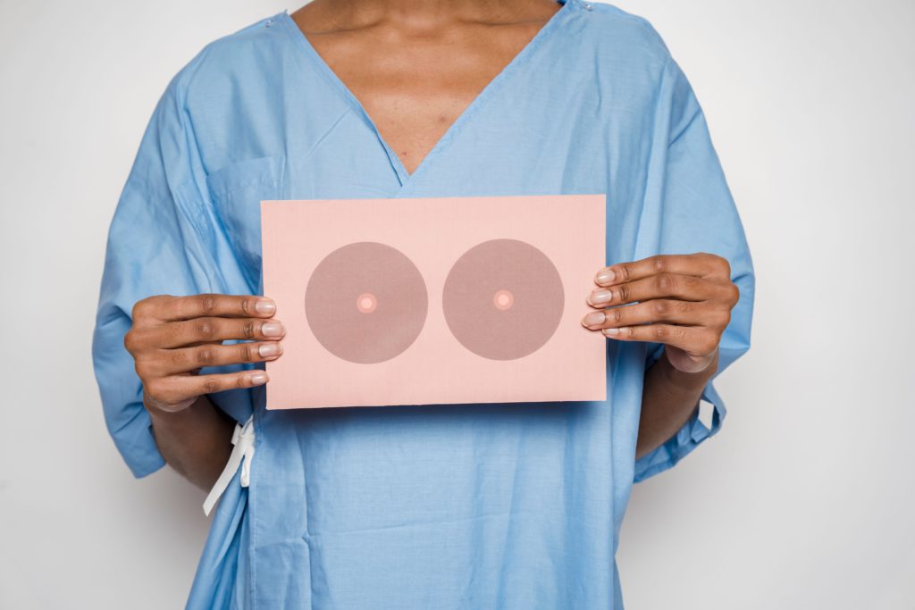
A mammogram is a medical imaging test that uses low-dose X-rays to produce detailed images of breast tissue. It is commonly used to screen for breast cancer before symptoms develop and may also be recommended when investigating changes such as a breast lump, unusual pain, nipple changes or other symptoms. The images are reviewed by appropriately trained medical professionals to identify areas that may require further assessment.
Mammography is the imaging technique used to create these breast images. During the examination, each breast is positioned on the mammography machine and briefly compressed between two plates. Compression spreads the breast tissue so clearer images can be obtained while keeping the required radiation dose low. The compression can feel uncomfortable for some people, but it generally lasts only a short time for each image.
Screening and Diagnostic Mammograms
Mammograms can be performed for different reasons. Screening mammography is used for people who do not have breast symptoms and aims to identify possible changes at an early stage.
Diagnostic mammography may be recommended when a person has symptoms or when an abnormality has been identified during another examination. Additional mammogram views or other imaging tests may be required to examine a particular area more closely.
How to Prepare
Most mammograms require relatively little preparation. However, following the imaging clinic’s instructions can make the examination easier.
Patients are commonly advised not to apply deodorant, powders, lotions or creams around the breasts and underarms on the day of the examination, because some products can interfere with the images.
Wearing a two-piece outfit can also be convenient because clothing from the waist up will generally need to be removed.
Tell the Imaging Team About Changes
Before the examination, patients should tell the radiographer about any breast changes or symptoms they have noticed. Previous breast surgery, implants and relevant medical history should also be discussed.
If previous mammogram images were taken at another imaging provider, having them available for comparison may help the radiologist assess whether an area has changed over time.
What Happens During the Test?
A specially trained radiographer positions the breast on the mammography unit. The breast is compressed while the X-ray image is taken, and several images are usually required.
The entire appointment takes longer than the actual X-ray exposure because time is needed for positioning and checking the images.
Checking and Understanding the Results
Patients should not attempt to interpret mammogram images themselves. A radiologist examines the images for changes or abnormalities and prepares a report for the referring healthcare professional or screening program.
An abnormal mammogram does not automatically mean that cancer is present. Additional mammogram views, ultrasound, MRI or a biopsy may sometimes be recommended to investigate a finding more thoroughly.
Follow Up Appropriately
Patients should follow the instructions provided by their healthcare professional or screening service regarding results and any further testing. They should also seek medical advice if they notice a new breast lump, nipple discharge, changes in breast shape or skin, or other unusual symptoms, even if a previous mammogram was normal.
Mammography is an important tool for examining breast tissue and detecting changes that may not be noticeable during a physical examination. Understanding how the test works, preparing appropriately and following up on results can help ensure breast imaging is used effectively as part of appropriate healthcare.